An Acute Thrombus Will Have Which of the Following Characteristics
An improved understanding of recanalization rates with thrombolysis and endovascular thrombectomy based on thrombus characteristics has spurred interest in new therapies for acute stroke. An acute thrombus will have which one of the following characteristics.

Characteristics Of Upper And Lower Extremity Deep Vein Thrombosis And Predictors Of Postthrombotic Syndrome In Children Sciencedirect
Superficial endothelial erosion leading to a clinically evident coronary thrombosis is most commonly seen in women and in diabetics with hypertriglyceridemia.

. Endothelial injury blood stasis and hypercoagulability. Many of the modalities used for detecting the presence of occlusive thrombus such as the hyperdense artery sign can also be used to describe thrombotic burden Another consideration in determining clot burden is the vessel segment in which thrombus is. Acute DVT often correlates with recent onset of symptoms.
Chronic DVT Acute DVT Dilated vein Non-compressible vein Occlusive thrombus with absent or near absent flow Hypoechoic thrombus same echo characteristics as blood Chronic DVT Vein not dilated Vein partially non-compressible Hyperechoic thrombus Stringy linear thrombus. Warmth in the skin of the affected leg. Embolic risk declines over time.
Absolute and relative density on CTA attenuation increase and void fraction. Acute thrombus in the deep veins is associated with the highest risk of thromboembolism. Deep vein thrombosis DVT is a condition of having a stationary blood clot in the deep veins of the arms or legs with a stationary blood clot being known as a thrombus.
Vein width at site of the thrombus is wider than the unaffected vein on the contralateral side ie dilated vessel and ultrasound echos are not prominent ie the clot is not echogenic. The manifestations and clinical significance of venous thrombus change over time. Venous thrombosis VT is a common clinical condition with a significant financial burden on the US healthcare system.
Thrombus characteristics included location clot burden score CBS length relative and absolute attenuation perviousness and distance from the internal carotid artery terminus to the thrombus. Location DT length volume absolute and relative attenuation on NCCT and thrombus perviousness measures. We have theorized that clots with stasis are longer.
The following speaker disclosures have the following relevant financial relationships with commercial interests. Bed rest or immobility for more than 4. Pain or tenderness in one or both legs which may occur only while standing or walking.
Thrombus characteristics included location clot burden score CBS length relative and absolute attenuation perviousness and distance from. What is the difference between a thrombus and a blood clot. Allen PharmD CACP Janssen Portola Speakers Bureau BMSPfizer Alliance Boehringer-Ingelheim Roche Consultant Geoffrey Barnes MD MSc PfizerBristol-Myers Squibb Janssen Acelis Connected Health Consultant.
Poorly attached to the vein wall b. Size or clot burden is an important factor as larger clots have been associated with poor response to therapy. Red or discolored skin in the affected leg.
3 This unique group of patients with SR was characterized by dilated left atria and decreased LAA. Poor attachment to wall d. We determined the following thrombus characteristics on baseline NCCT and CTA imaging.
A blood clot is any coagulated mass of blood but a thrombus is a blood clot that occurs specifically in a blood vessel or the heart. In venous duplex scanning acute DVT has all of the following characteristics except. Classic Duplex Findings in Acute vs.
Acute DVT in which the. Acute venous thrombosis confirmed by duplex ultrasound has the following characteristics. We therefore explored the relationship between thrombus imaging characteristics on noncontrast computed tomography NCCT and magnetic resonance imaging MRI with clot length and pial collaterals on baseline computed tomography angiography CTA.
About 70 of cases of acute coronary thrombosis involve a disrupted atherosclerotic plaque and in the remaining 30 there is only superficial intimal injury at the site of thrombus formation. All characteristics were assessed on thin-slice 25 mm noncontrast computed. Left ventricular LV thrombus development following acute myocardial infarction is driven by the elements of Virchows triad.
13 Location of the thrombus M1 M2 A1 A2 ICA or ICA-T was determined on CTA. It will be rigid and nondeformable d. If you have any of the following DVT symptoms especially if they occur suddenly call your doctor immediately.
Location distance from ICA terminus to thrombus length volume absolute andrelativedensityonNCCTandperviousnessAssociationsofthrombuscharacteristicswithoutcomewereestimatedwithunivariable and multivariable ordinal logistic regression as an OR for a shift toward better outcome on the. In univariate analyses the LV thrombus group was more likely to have atrial fibrillation chronic or acute heart failure with reduced ejection fraction left heart valvular disease left ventricular aneurysm history of chronic or acute pulmonary embolism or deep venous thrombosis and anterior or anterolateral myocardial infarction 77 vs. Despite its prevalence the diagnosis and management VT remains rudimentary 1-4Anticoagulation and thrombolytic therapies are the mainstay of VT treatment despite a rapid decrease in their effectiveness as thrombus.
As the thrombus ages it becomes less friable increasingly fibrotic and more firmly attached to the vein wall. Both viable myocardium and thrombus can appear relatively dark and may be difficult to distinguish from one another. We assessed the following thrombus characteristics.
Swelling in one or both legs. On conventional DE-CMRwhich is tailored to null viable myocardium typical inversion time 200-300 msecthrombus appears grey or etched viable myocardium black and infarcted myocardium white. Each of these components further serves as a therapeutic target in the treatment and prevention of left ventricular thrombus following acute myocardial infarction.
Background and Objectives New imaging techniques have advanced our ability to capture thrombus characteristics and burden in real time. In a recently published study of patients with acute 24 hours ischemic neurological events LA thrombus was detected in 6 of 583 patients with SR 3 patients had mitral stenosis and the other 3 had aortic stenosis coronary artery disease and cardiomyopathy.

Incidence And Risk Factors For Deep Vein Thrombosis After Radiofrequency And Laser Ablation Of The Lower Extremity Veins Annals Of Vascular Surgery

Thrombus Composition And Etiology Of Stroke Download Scientific Diagram
Histologic Assessment Of Lower Extremity Deep Vein Thrombus From Patients Undergoing Percutaneous Mechanical Thrombectomy Journal Of Vascular Surgery Venous And Lymphatic Disorders

Shape Of The Thrombus A Concentric Thrombus The Thrombus Causes Total Download Scientific Diagram

Clinical Significance Of Hepatosplenic Thrombosis In Vaccine Induced Immune Thrombotic Thrombocytopenia After Chadox1 Ncov 19 Vaccination International Journal Of Infectious Diseases

Shape Of The Thrombus A Concentric Thrombus The Thrombus Causes Total Download Scientific Diagram

Introduction To Coronary Artery Disease Ischemic Heart Disease Use Of Ecg Ecg Echo In 2021 Ischemic Heart Disease Coronary Artery Disease Coronary Arteries
Interplay Between Inflammation And Thrombosis In Cardiovascular Pathology Nature Reviews Cardiology

Venous Sinus Thrombosis In A Preterm Male Neonate With Elevated Anti Phospholipid Antibodies A Case Report Thrombosis Sinusitis Neonatal
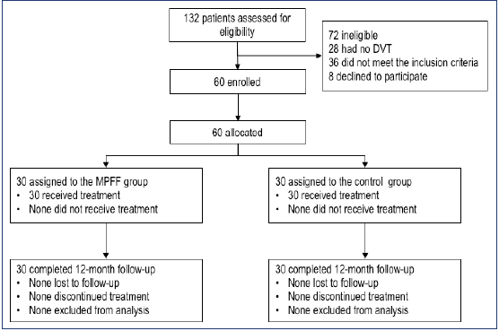
Micronized Purified Flavonoid Fraction In Adjunction To Rivaroxaban Improves Outcomes Of Popliteal Femoral Deep Vein Thrombosis At 12 Month Follow Up Servier Phlebolymphologyservier Phlebolymphology

Cord Sign Radiology Reference Article Radiopaedia Org Radiology Thrombosis Brain Images

Critical Review Of Large Animal Models For Central Deep Venous Thrombosis European Journal Of Vascular And Endovascular Surgery
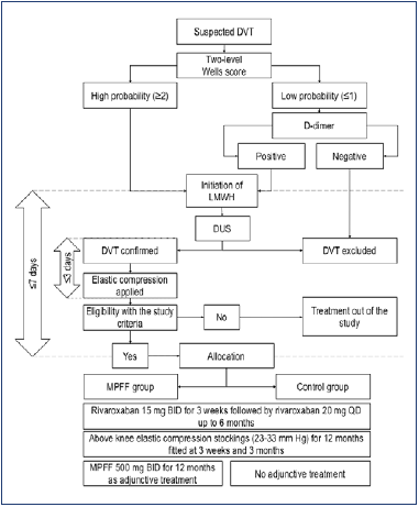
Micronized Purified Flavonoid Fraction In Adjunction To Rivaroxaban Improves Outcomes Of Popliteal Femoral Deep Vein Thrombosis At 12 Month Follow Up Servier Phlebolymphologyservier Phlebolymphology

Cornary Artey Disease Coronary Artery Disease Ischemic Heart Disease Myocardial Infarction

Thrombus Composition And Etiology Of Stroke Download Scientific Diagram

Imaging Of Cerebral Venous Thrombosis Clinical Radiology

Editor S Choice European Society For Vascular Surgery Esvs 2021 Clinical Practice Guidelines On The Management Of Venous Thrombosis European Journal Of Vascular And Endovascular Surgery

Shape Of The Thrombus A Concentric Thrombus The Thrombus Causes Total Download Scientific Diagram

Zakhem B Fernandez Ortiz A Alfonso F Optical Coherence Tomography Findings During Evolving Optical Coherence Tomography Thrombosis Cardiac Catheterization


Comments
Post a Comment